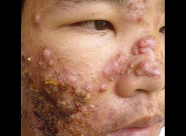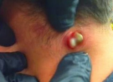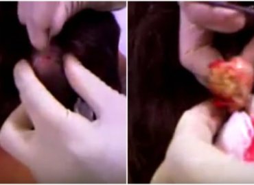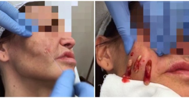
Granulomas are a delayed, poorly understood and distressing complication of dermal filler injections. Histologically, they represent a foreign body type of reaction with giant cells and macrophages infiltrating tissues. Granulomas are classified into three types: cystic, nodular and sclerosing, which clinically present as red, firm papules, nodules or plaques occurring months or years after filler injections. Filler dependent factors such as volume and particle size, as well as the presence of biofilm, have been suggested as possible causes.
 Treatment is often empirical, hence good differential diagnosis is essential in choosing the right treatment pathway. Overcorrection using dermal fillers, hypersensitivity reaction and infections ought to be considered before embarking on treatment, which consists of antibiotics and hyaluronidase in the first instance, followed by intralesional or systemic steroids. Surgical excision is recommended as the last resort.
 Investigations that may be of assistance in making the diagnosis and assist with the management include blood tests such as: white blood count (WBC) and C-reactive protein (CRP). Out of the imaging methods available, the use of ultrasound (USS) is most useful. Biopsy and histology offer confirmatory diagnosis. Culture is often unhelpful as a negative result does not exclude possibility of the presence of biofilm. Counselling the patient and adopting preventative measures such appropriate filler choice and prevention of infection should be part of every case of dermal filler injections.
 Granulomas following dermal filler injections are challenging for aesthetic practitioners to manage and can be distressing to the individuals affected by them.1,2 Granulomas can develop months or years after deposition of the dermal filler in the dermis and may occur after both permanent and semi-permanent dermal filler use, including hyaluronic acid (HA), bovine collagen, silicone, paraffin, polyacrylamide gel, poly-lactic acid microspheres and calcium hydroxyapatite.1-8 Frequency of granuloma occurrence has been reported as 0.02-0.4% after HA and 0.04%-0.3% after bovine collagen injections.
 Granuloma formation is a non-allergic, chronic inflammatory response, characterised by foreign body types of reaction in the dermis following injection of dermal filler or other foreign material. A granuloma is defined as a tumour composed of immune cells such as macrophages, activated and fused into multinucleated giant cells, consisting of more than 20 nuclei and arranged in an irregular and random way.
Formation of granuloma occurs in stages involving:
- Protein absorption
- Macrophage adhesion
- Macrophage fusion
- Crosstalk
As the neutrophil infiltration and adsorption of host proteins to the foreign material occur, monocytes circulating in the blood migrate to the surrounding tissues and differentiate into macrophages. Where the particle volume is greater than the macrophage volume, macrophages aggregate, forming giant cells and secrete factors, which activate fibroblasts, influencing the development of fibrous capsule around the foreign body material and formation of the foreign body giant cell (FBGC).
 It is not fully understood why small filler particles or silicone fluid trigger granuloma formation, both of which are easily phagocytosed by macrophages.One hypothesis that was put forward by Lemperle et al in 2006 suggests that macrophages act as memory cells and remember small phagocytosed particles. Triggered by systemic infection, they initiate FBGC formation and the development of granuloma.The authors were of the opinion that volume, purity and physical characteristics of the injected dermal filler such as particle size, smoothness, charge and hydrophilicity play a role in this process.
 Bentkover hypothesised that the main cause of FBGC reaction is the size of the filler particles, which prevents them from being phagocytosed.Granulomas have been linked to biofilms, which are defined as a structured community of microorganisms encapsulated within a self-developed, polymeric matrix, irreversibly adherent to a living or inert surface.In 2007, Christiansen hypothesised that biofilms form when bacteria is introduced during filler injections or are seeded in the filler during bacteriaemic episodes.Once present, they remain dormant for months or years on the surface of the filler and become a target of a delayed immune response, resulting in granuloma formulation. Many biofilms are almost impossible to culture using current microbiology culture technology.They live in a quiescent state, resulting in a low-grade infection associated with low-grade host response. Activation of biofilms may be triggered by dental manipulation, trauma or other factors, leading to local or systemic infection, as well as granulomatous, inflammatory response. Biofilm populations can shift from active to dormant depending on exogenous threats. When bacterial proteins turn off cell metabolism and the cell becomes dormant, it becomes antibiotic resistant, as well as difficult, if not impossible, to culture.
 Patients with chronic sinusitis, chronic dental problems, or other infections may have a greater tendency to develop an infection after a filler is injected in the periorbital area or central face. These patients may also be prone to formation of a biofilm around or in the implant, caused by injection trauma around the site of a previous filler injection. Many problems that were previously assumed to be foreign body granulomas or allergic reactions, on the basis of negative bacterial cultures, are now thought to be due to biofilms.
Diagnosis and treatment of dermal filler granulomas
Diagnosis and management of dermal filler granulomas is complex. In order to offer satisfactory treatment and resolution, it is very important to take good medical and aesthetic history and thoroughly examine the patient. Investigations may assist with making correct diagnosis although, in reality, they are rarely carried out and treatment is empirical. It is therefore essential to understand the pathophysiology of granulomas and differentiate them from other causes of nodular lesions caused by dermal fillers, as their management differs depending on the presence or absence of inflammatory features.
 Conclusion
The filler is a filling technique that removes wrinkles and blemishes, however, it has side effects that appear over time: filler granulomas. These are chronic inflammations that can cause discomfort and break the harmony of the face. The remedies that are used to combat this problem have as main protagonist the laser. The most used method is called ILT, a treatment that allows to use the heat to fight against the filler. Heat affects molecules by moving them from the solid state to the liquid, so that it can be removed later. An additional advantage of this technique is the stimulation of the production of collagen by the tissues, which appear more toned and elastic.