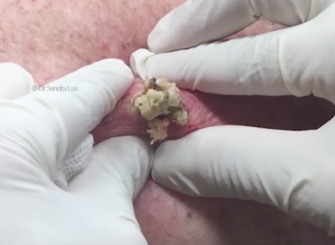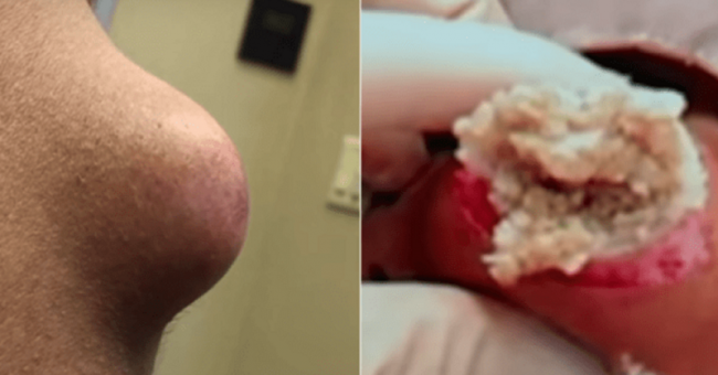
An epidermoid cyst (Epidermal Inclusion cyst, Infundibular cyst), is a benign growth commonly found in the skin and typically appears on the face, neck or trunk, but can occur anywhere on the body. Another name used is “sebacous cyst” but this is actually an antiquated misnomer, and is not a term used by dermatologists. They are also the most common type of cutaneous cysts. Epidermoid cysts result from the reproduction of epidermal cells within a confined space of the dermis. The pasty contents are mostly composed of macerated keratin (wet skin cells), which creates this “cheesy” consistency, and there can be a pungent odor.
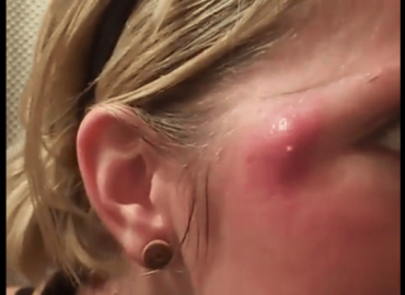
 An epidermoid cyst may have no symptoms and are typically harmless. Usually people seek removal but they don’t like the appearance of these bumps, or the cyst has ruptured or been inflamed or “infected” in the past. Rupture is associated with sudden redness, pan, swelling, and local heat, and can lead to abscess formation. Also, a history of inflammation, often increases scar tissue in the area, makes the cyst more firmly adherent to surrounding skin, and makes it more difficult to remove. Surgical excision is curative, but the complete cyst removal including the entire cyst sac and contents need to be removed to ensure that the cyst won’t reoccur.
 Physical Examination
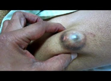
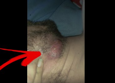
Epidermoid cysts appear as flesh–colored-to-yellowish, firm, round nodules of variable size. A central pore or punctum may be present. Note the image below.
Reportedly, epidermoid cysts are most common (in descending order of frequency) on the face, trunk, neck, extremities, and scalp. Rare cases of epidermoid cysts occurring in bone, breast, and various intracranial locations have been reported. Epidermoid cysts in the breast and genital area are not uncommon in the general population. The ocular and oral mucosae can also be affected and, cysts have been reported on the palpebral conjunctivae, lips, buccal mucosa, tongue, and uvula.
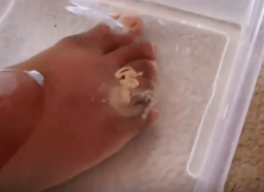
Epidermoid cysts can manifest in various ways on the extremities. Epidermoid cysts on the distal portions of the digits may extend into the terminal phalanx. These lesions may produce changes in the nails such as pincer nails, erythema, edema, tenderness, and pain.
Causes
Epidermoid cysts likely form by several mechanisms. They may result from the sequestration of epidermal rests during embryonic life, occlusion of the pilosebaceous unit, or traumatic or surgical implantation of epithelial elements. HPV infection, ultraviolet exposure, and eccrine duct occlusion may be additional factors in the development of palmoplantar epidermoid cysts. HPV has also been identified in nonpalmoplantar epidermoid cysts.
Congenital epidermoid cysts of the anterior fontanelle or those that are orogenital in location presumably result from sequestration or trapping of epidermal rests along embryonic fusion planes during development. Lip and lingual lesions may be related to aberrant fusion of the branchial arches, while genital lesions may result from improper closure of the genital folds.
Any benign or malignant process affecting or growing near the pilosebaceous unit may lead to occlusion or impingement of the follicular ostia with subsequent formation of a cyst. Cysts with an acneiform distribution are likely the result of follicular occlusion. In elderly persons, accumulated sun damage can injure the pilosebaceous unit, causing abnormalities such as comedonal plugging and hypercornification, both of which can eventuate in cyst formation.This condition is referred to as Favre-Racouchot syndrome.
 True epidermal inclusion cysts result from the implantation of epithelial elements in the dermis. Certain injuries, especially of the crushing type, have been associated with subungual or terminal phalanx epidermoid cysts. A crush injury sustained from slamming a car door on a digit is frequently reported. Any surgical procedure can theoretically result in epidermoid cysts. Reports describe the formation of multiple epidermoid cysts after rhinoplasty, breast augmentation, and liposuction.The use of dermal grafts, myocutaneous grafts, and needle biopsies has also been associated with the formation of epidermoid cysts.
Certain hereditary syndromes are associated with epidermoid cysts. Such syndromes include Gardner syndrome,basal cell nevus syndrome,and pachyonychia congenita.In addition, idiopathic scrotal calcinosis may actually represent an end stage of dystrophic calcification of epidermoid cysts.